Imaging Colonic Surface Topography With Photometric Stereo Endoscopy
Durr NJ, Parot V, Traverso G, Puricelli WP, Vakoc BJ, Nishioka NS, Gonzalez G.
DDW 2014 Gastrointestinal Endoscopy 79:5 2014.
DOI: 10.1016/j.gie.2014.02.676
Abstract
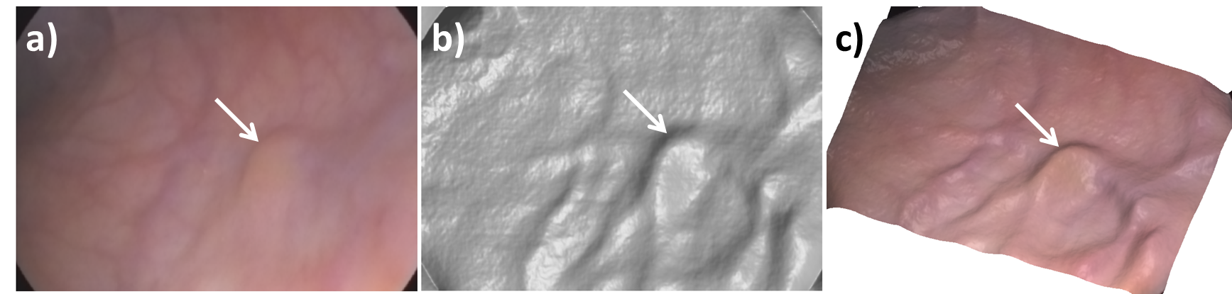
Colorectal lesions are usually classified by their characteristic three-dimensional morphology. Conventional colonoscopy captures the color and reflectivity contrast of the mucosal surface, but the surface topography can only be inferred by the endoscopist through indirect cues. Consequently, flat lesions with subtle color contrast can be difficult to identify. In a previous ex-vivo study, we demonstrated that photometric stereo endoscopy (PSE) can capture the high-frequency surface topography of colon tissue along with the conventional white-light image using a standard endoscope.
Aims
The aim of this research was to evaluate the capability of PSE to capture relevant lesion topography in the human rectum in-vivo.
Methods
The PSE endoscope consists of a commercial gastroscope with a custom illumination cap attached to the distal end. The illumination cap holds four fiber optic light guides that strobe synchronously with the frame rate of the endoscope image sensor. The assembled system has the same outer diameter as a conventional colonoscope (14 mm). The acquired images are processed in real-time to display a conventional white-light image to the endoscopist. The surface topography is then extracted from the recorded movie with post-processing.
Results
To date we have successfully imaged the rectum of 8 subjects using PSE. When imaging obliquely to the mucosa surface, PSE reconstructs detailed topography that correlates with the expected shape from observing the conventional image. Large blood vessels and diminutive lesions are reconstructed as elevations in the tissue surface. The captured topography can be used to enhance the contrast of elevated or recessed features. When imaging perpendicular to the tissue surface, the current PSE algorithm generates topographical artifacts from specular reflections.
Conclusions
Our initial experience with PSE has shown that the technique is capable of obtaining high quality topographic images of blood vessels, lesions, and tissue folds in the human rectum. These results suggest that PSE is a promising technique for increasing lesion contrast during endoscopic procedures.