Back-illumination interference tomography for imaging weak scattering in thick tissues
McKay GN, Mertz J, Durr NJ.
Optica 12:12 2025 [Journal Link] [Preprint]
Abstract
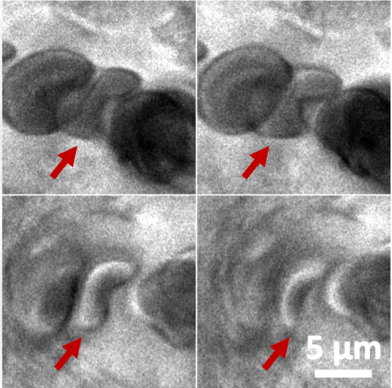
Biological tissues are composed of discrete compartments of biochemical media that often exhibit subtle differences in refractive index. Light propagating through these compartments partially diffracts in a forward direction with a pi/2 phase shift. We introduce a microscopy technique to image this scattering signal in thick tissues, called back-illumination interference tomography (BIT). An incoherent source is demagnified and imaged past the focal plane of a high numerical aperture objective lens, producing a small, semicoherent source of backscattered light. This backscattered light undergoes a phase inversion over the narrow depth of field of the microscope, providing interference contrast to weakly scattering objects at the focal plane. BIT offers a fundamentally different source of contrast to conventional illumination and oblique back-illumination microscopy. Compared to these techniques, we show that BIT improves contrast to blood cells in vitro in microfluidic chambers and in vivo in a human capillary. Finally, we apply BIT to unstained, unlabeled bulk human tissue ex vivo and compare side-by-side to adjacent frozen sections stained with Hematoxylin and Eosin. These results demonstrate the potential of BIT to provide high resolution, high speed, 3D imaging of unprocessed biological tissues.
