First-in-human pilot study of a spatial frequency domain oxygenation imaging system
Gioux S, Mazhar A, Lee BT, Lin SJ, Tobias AM, Cuccia DJ, Stockdale A, Oketokoun R, Ashitate Y, Kelly E, Weinmann M, Durr NJ, Moffitt LA, Durkin AJ, Tromberg BJ, Frangioni JV.
Journal of Biomedical Optics 16:8 2011.
DOI: 10.1117/1.3614566
Abstract
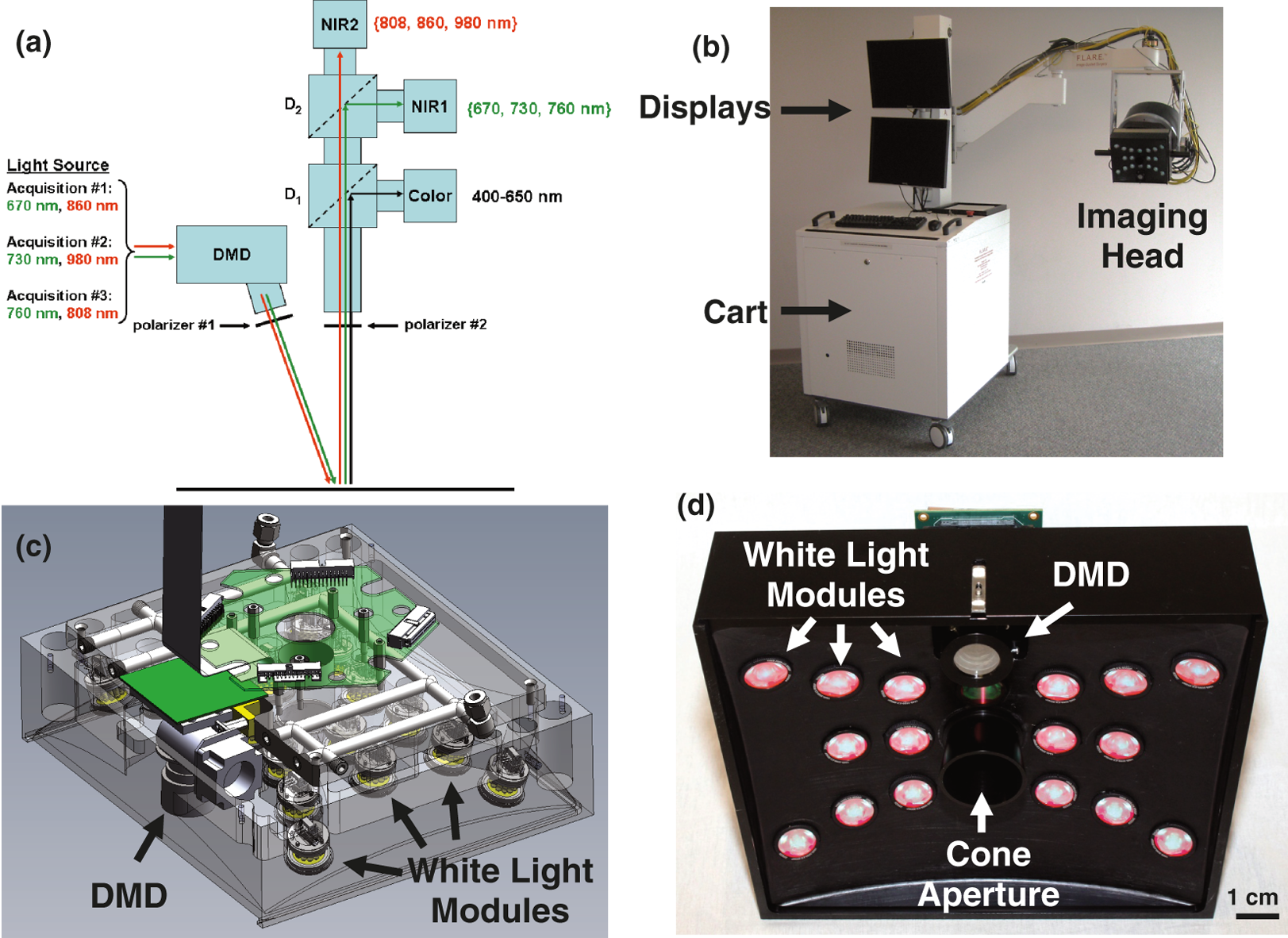
Oxygenation measurements are widely used in patient care. However, most clinically available instruments currently consist of contact probes that only provide global monitoring of the patient (e.g., pulse oximetry probes) or local monitoring of small areas (e.g., spectroscopy-based probes). Visualization of oxygenation over large areas of tissue, without a priori knowledge of the location of defects, has the potential to improve patient management in many surgical and critical care applications. In this study, we present a clinically compatible multispectral spatial frequency domain imaging (SFDI) system optimized for surgical oxygenation imaging. This system was used to image tissue oxygenation over a large area (16×12 cm) and was validated during preclinical studies by comparing results obtained with an FDA-approved clinical oxygenation probe. Skin flap, bowel, and liver vascular occlusion experiments were performed on Yorkshire pigs and demonstrated that over the course of the experiment, relative changes in oxygen saturation measured using SFDI had an accuracy within 10% of those made using the FDA-approved device. Finally, the new SFDI system was translated to the clinic in a first-in-human pilot study that imaged skin flap oxygenation during reconstructive breast surgery. Overall, this study lays the foundation for clinical translation of endogenous contrast imaging using SFDI.