Photometric Stereo Endoscopy
Parot V, Lim D, Gonzalez G, Traverso CG, Nishioka NS, Vakoc B, Durr NJ
Journal of Biomedical Optics 18:7 2013.
DOI: 10.1117/1.JBO.18.7.076017
Abstract
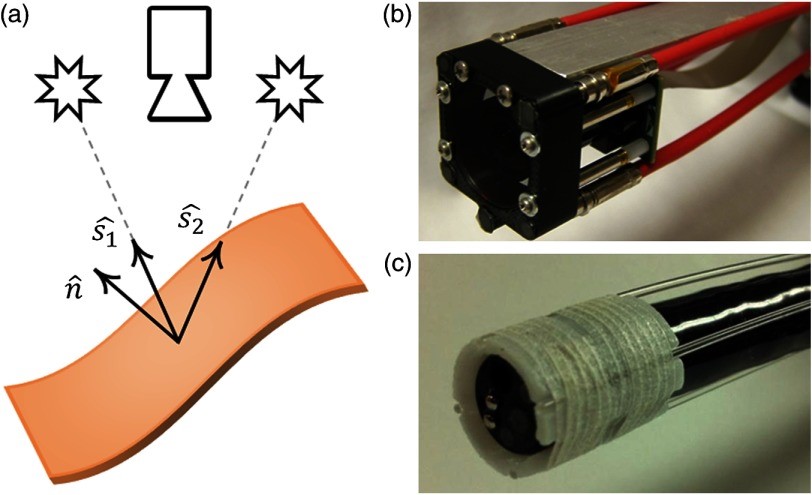
While color video endoscopy has enabled wide-field examination of the gastrointestinal tract, it often misses or incorrectly classifies lesions. Many of these missed lesions exhibit characteristic three-dimensional surface topographies. An endoscopic system that adds topographical measurements to conventional color imagery could therefore increase lesion detection and improve classification accuracy. We introduce photometric stereo endoscopy (PSE), a technique which allows high spatial frequency components of surface topography to be acquired simultaneously with conventional two-dimensional color imagery. We implement this technique in an endoscopic form factor and demonstrate that it can acquire the topography of small features with complex geometries and heterogeneous optical properties. PSE imaging of ex vivo human gastrointestinal tissue shows that surface topography measurements enable differentiation of abnormal shapes from surrounding normal tissue. Together, these results confirm that the topographical measurements can be obtained with relatively simple hardware in an endoscopic form factor, and suggest the potential of PSE to improve lesion detection and classification in gastrointestinal imaging.